Connect with us

The Conversation

Women are at a higher risk of dying from heart disease − in part because doctors don’t take major sex and gender differences into account

Published

on

theconversation.com – Amy Huebschmann, Professor of Medicine, University of Colorado Anschutz Medical Campus – 2024-10-22 07:45:00

Rates of heart disease and cardiac events in women are often underestimated.

eternalcreative/iStock via Getty Images

Amy Huebschmann, University of Colorado Anschutz Medical Campus and Judith Regensteiner, University of Colorado Anschutz Medical Campus

A simple difference in the genetic code – two X chromosomes versus one X chromosome and one Y chromosome – can lead to major differences in heart disease. It turns out that these genetic differences influence more than just sex organs and sex assigned at birth – they fundamentally alter the way cardiovascular disease develops and presents.

While sex influences the mechanisms behind how cardiovascular disease develops, gender plays a role in how health care providers recognize and manage it. Sex refers to biological characteristics such as genetics, hormones, anatomy and physiology, while gender refers to social, psychological and cultural constructs. Women are more likely to die after a first heart attack or stroke than men. Women are also more likely to have additional or different heart attack symptoms that go beyond chest pain, such as nausea, jaw pain, dizziness and fatigue. It is often difficult to fully disentangle the influences of sex on cardiovascular disease outcomes versus the influences of gender.

While women who haven’t entered menopause have a lower risk of cardiovascular disease than men, their cardiovascular risk accelerates dramatically after menopause. In addition, if a woman has Type 2 diabetes, her risk of heart attack accelerates to be equivalent to that of men, even if the woman with diabetes has not yet gone through menopause. Further data is needed to better understand differences in cardiovascular disease risk among nonbinary and transgender patients.

Despite these differences, one key thing is the same: Heart attack, stroke and other forms of cardiovascular disease are the leading cause of death for all people, regardless of sex or gender.

We are researchers who study women’s health and the way cardiovascular disease develops and presents differently in women and men. Our work has identified a crucial need to update medical guidelines with more sex-specific approaches to diagnosis and treatment in order to improve health outcomes for all.

Gender differences in heart disease

The reasons behind sex and gender differences in cardiovascular disease are not completely known. Nor are the distinct biological effects of sex, such as hormonal and genetic factors, versus gender, such as social, cultural and psychological factors, clearly differentiated.

What researchers do know is that the accumulated evidence of what good heart care should look like for women compared with men has as many holes in it as Swiss cheese. Medical evidence for treating cardiovascular disease often comes from trials that excluded women, since women for the most part weren’t included in scientific research until the NIH Revitalization Act of 1993. For example, current guidelines to treat cardiovascular risk factors such as high blood pressure are based primarily on data from men. This is despite evidence that differences in the way that cardiovascular disease develops leads women to experience cardiovascular disease differently.

Clinician holding stethoscope against a patient's chest

Gender biases in health care influence the kind of tests and attention that women receive.
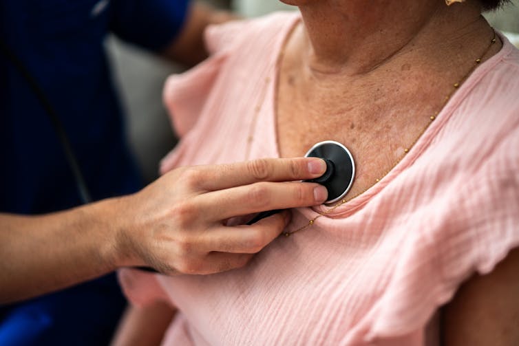
FG Trade Latin/E+ via Getty Images

In addition to sex differences, implicit gender biases among providers and gendered social norms among patients lead clinicians to underestimate the risk of cardiac events in women compared with men. These biases play a role in why women are more likely than men to die from cardiac events. For example, for patients with symptoms that are borderline for cardiovascular disease, clinicians tend to be more aggressive in ordering artery imaging for men than for women. One study linked this tendency to order less aggressive tests for women partly to a gender bias that men are more open than women to taking risks.

In a study of about 3,000 patients with a recent heart attack, women were less likely than men to think that their heart attack symptoms were due to a heart condition. Additionally, most women do not know that cardiovascular disease is the No. 1 cause of death among women. Overall, women’s misperceptions of their own risk may hold them back from getting a doctor to check out possible symptoms of a heart attack or stroke.

These issues are further exacerbated for women of color. Lack of access to health care and additional challenges drive health disparities among underrepresented racial and ethnic minority populations.

Sex difference in heart disease

Cardiovascular disease physically looks different for women and men, specifically in the plaque buildup on artery walls that contributes to illness.

Women have fewer cholesterol crystals and fewer calcium deposits in their artery plaque than men do. Physiological differences in the smallest blood vessels feeding the heart also play a role in cardiovascular outcomes.

Women are more likely than men to have cardiovascular disease that presents as multiple narrowed arteries that are not fully “clogged,” resulting in chest pain because blood flow can’t ratchet up enough to meet higher oxygen demands with exercise, much like a low-flow showerhead. When chest pain presents in this way, doctors call this condition ischemia and no obstructive coronary arteries. In comparison, men are more likely to have a “clogged” artery in a concentrated area that can be opened up with a stent or with cardiac bypass surgery. Options for multiple narrowed arteries have lagged behind treatment options for typical “clogged” arteries, which puts women at a disadvantage.

In addition, in the early stages of a heart attack, the levels of blood markers that indicate damage to the heart are lower in women than in men. This can lead to more missed diagnoses of coronary artery disease in women compared with men.

The reasons for these differences are not fully clear. Some potential factors include differences in artery plaque composition that make men’s plaque more likely to rupture or burst and women’s plaque more likely to erode. Women also have lower heart mass and smaller arteries than men even after taking body size into consideration.

Reducing sex disparities

Too often, women with symptoms of cardiovascular disease are sent away from doctor’s offices because of gender biases that “women don’t get heart disease.”

Considering how symptoms of cardiovascular disease vary by sex and gender could help doctors better care for all patients.

One way that the rubber is meeting the road is with regard to better approaches to diagnosing heart attacks for women and men. Specifically, when diagnosing heart attacks, using sex-specific cutoffs for blood tests that measure heart damage – called high-sensitivity troponin tests – can improve their accuracy, decreasing missed diagnoses, or false negatives, in women while also decreasing overdiagnoses, or false positives, in men.

Our research laboratory’s leaders,collaborators and other internationally recognized research colleagues – some of whom partner with our Ludeman Family Center for Women’s Health Research on the University of Colorado Anschutz Medical Campus – will continue this important work to close this gap between the sexes in health care. Research in this field is critical to shine a light on ways clinicians can better address sex-specific symptoms and to bring forward more tailored treatments.

The Biden administration’s recent executive order to advance women’s health research is paving the way for research to go beyond just understanding what causes sex differences in cardiovascular disease. Developing and testing right-sized approaches to care for each patient can help achieve better health for all.The Conversation

Amy Huebschmann, Professor of Medicine, University of Colorado Anschutz Medical Campus and Judith Regensteiner, Professor of Medicine, University of Colorado Anschutz Medical Campus

This article is republished from The Conversation under a Creative Commons license. Read the original article.

Read More

The post Women are at a higher risk of dying from heart disease − in part because doctors don’t take major sex and gender differences into account appeared first on theconversation.com

The Conversation

Transplanting insulin-making cells to treat Type 1 diabetes is challenging − but stem cells offer a potential improvement

Published

on

theconversation.com – Vinny Negi, Research Scientist in Endocrinology and Metabolism, University of Pittsburgh – 2024-11-20 07:36:00

The islets of Langerhans play a crucial role in blood sugar regulation.
Fayette A Reynolds/Berkshire Community College Bioscience Image Library via Flickr

Vinny Negi, University of Pittsburgh

Diabetes develops when the body fails to manage its blood glucose levels. One form of diabetes causes the body to not produce insulin at all. Called Type 1 diabetes, or T1D, this autoimmune disease happens when the body’s defense system mistakes its own insulin-producing cells as foreign and kills them. On average, T1D can lead patients to lose an average of 32 years of healthy life.

Current treatment for T1D involves lifelong insulin injections. While effective, patients taking insulin risk developing low blood glucose levels, which can cause symptoms such as shakiness, irritability, hunger, confusion and dizziness. Severe cases can result in seizures or unconsciousness. Real-time blood glucose monitors and injection devices can help avoid low blood sugar levels by controlling insulin release, but they don’t work for some patients.

For these patients, a treatment called islet transplantation can help better control blood glucose by giving them both new insulin-producing cells as well as cells that prevent glucose levels from falling too low. However, it is limited by donor availability and the need to use immunosuppressive drugs. Only about 10% of T1D patients are eligible for islet transplants.

In my work as a diabetes researcher, my colleagues and I have found that making islets from stem cells can help overcome transplantation challenges.

History of islet transplantation

Islet transplantation for Type 1 diabetes was FDA approved in 2023 after more than a century of investigation.

Insulin-producing cells, also called beta cells, are located in regions of the pancreas called islets of Langerhans. They are present in clusters of cells that produce other hormones involved in metabolism, such as glucagon, which increases blood glucose levels; somatostatin, which inhibits insulin and glucagon; and ghrelin, which signals hunger. Anatomist Paul Langerhans discovered islets in 1869 while studying the microscopic anatomy of the pancreas, observing that these cell clusters stained distinctly from other cells.

The road to islet transplantation has faced many hurdles since pathologist Gustave-Édouard Laguesse first speculated about the role islets play in hormone production in the late 19th century. In 1893, researchers attempted to treat a 13-year-old boy dying of diabetes with a sheep pancreas transplant. While they saw a slight improvement in blood glucose levels, the boy died three days after the procedure.

Microscopy image of oblong blob of yellow and pink cells surrounded by violet cells
The islets of Langerhans, located in the pancreas and colored yellow here, secrete hormones such as insulin and glucagon.
Steve Gschmeissner/Science Photo Library via Getty Images

Interest in islet transplantation was renewed in 1972, when scientist Paul E. Lacy successfully transplanted islets in a diabetic rat. After that, many research groups tried islet transplantation in people, with no or limited success.

In 1999, transplant surgeon James Shapiro and his team successfully transplanted islets in seven patients in Edmonton, Canada, by transplanting a large number of islets from two to three donors at once and using immunosuppressive drugs. Through the Edmonton protocol, these patients were able to manage their diabetes without insulin for a year. By 2012, over 1,800 patients underwent islet transplants based on this technique, and about 90% survived through seven years of follow-up. The first FDA-approved islet transplant therapy is based on the Edmonton protocol.

Stem cells as a source of islets

Islet transplantation is now considered a minor surgery, where islets are injected into a vein in the liver using a catheter. As simple as it may seem, there are many challenges associated with the procedure, including its high cost and a limited availability of donor islets. Transplantation also requires lifelong use of immunosuppressive drugs that allow the foreign islets to live and function in the body. But the use of immunosuppressants also increases the risk of other infections.

To overcome these challenges, researchers are looking into using stem cells to create an unlimited source of islets.

There are two kinds of stem cells scientists are using for islet transplants: embryonic stem cells, or ESCs, and induced pluripotent stem cells, or iPSCs. Both types can mature into islets in the lab.

Each has benefits and drawbacks.

There are ethical concerns regarding ESCs, since they are obtained from dead human embryos. Transplanting ESCs would still require immunosuppressive drugs, limiting their use. Thus, researchers are working to either encapsulate or make mutations in ESC islets to protect them from the body’s immune system.

Conversely, iPSCs are obtained from skin, blood or fat cells of the patient undergoing transplantation. Since the transplant involves the patient’s own cells, it bypasses the need for immunosuppressive drugs. But the cost of generating iPSC islets for each patient is a major barrier.

A long life with Type 1 diabetes is possible.

Stem cell islet challenges

While iPSCs could theoretically avoid the need for immunosuppressive drugs, this method still needs to be tested in the clinic.

T1D patients who have genetic mutations causing the disease currently cannot use iPSC islets, since the cells that would be taken to create stem cells may also carry the same disease-causing mutation of their islet cells. Many available gene-editing tools could potentially remove those mutations and generate functional iPSC islets.

In addition to the challenge of genetic tweaking, price is a major issue for islet transplantation. Transplanting islets made from stem cells is more expensive than insulin therapy because of higher manufacturing costs. Efforts to scale up the process and make it more cost effective include creating biobanks for iPSC matching. This would allow iPSC islets to be used for more than one patient, reducing costs by avoiding the need to generate freshly modified islets for each patient. Embryonic stem cell islets have a similar advantage, as the same batch of cells can be used for all patients.

There is also a risk of tumors forming from these stem cell islets after transplantation. So far, lab studies on rodents and clinical trials in people have rarely shown any cancer. This suggests the chances of these cells forming a tumor are low.

That being said, many rounds of research and development are required before stem cell islets can be used in the clinic. It is a laborious trek, but I believe a few more optimizations can help researchers beat diabetes and save lives.

Article updated to clarify that Type 1 diabetes causes the body to not produce insulin.The Conversation

Vinny Negi, Research Scientist in Endocrinology and Metabolism, University of Pittsburgh

This article is republished from The Conversation under a Creative Commons license. Read the original article.

Read More

The post Transplanting insulin-making cells to treat Type 1 diabetes is challenging − but stem cells offer a potential improvement appeared first on theconversation.com

Continue Reading

The Conversation

Should I worry about mold growing in my home?

Published

on

theconversation.com – Nicholas Money, Professor of Biology, Miami University – 2024-11-20 07:36:00

Mold growths are common in homes, and unless the damage is widespread, they usually aren’t harmful.

AP Photo/Matt Rourke

Nicholas Money, Miami University

Mold growth in your home can be unsettling. Blackened spots and dusty patches on the walls are signs that something is amiss, but it is important to distinguish between mold growth that is a nuisance and mold growth that may be harmful.

There are more than 1 million species of fungi. Some are used to produce important medications. Others can cause life-threatening infections when they grow in the body.

Microscopic fungi that grow in homes are a problem because they can trigger asthma and other allergies. In my work as a fungal biologist, however, I have yet to encounter robust scientific evidence to support claims that indoor molds are responsible for other serious illnesses.

What are molds?

Molds are microscopic fungi that grow on everything. This may sound like an exaggeration, but pick any material and a mold will be there, from the leaves on your houseplant to the grain in your pantry and every pinch of soil on the ground. They form splotches on the outside of buildings, grow in crevices on concrete paths and roads, and even live peacefully on our bodies.

Molds are important players in life on Earth. They’re great recyclers that fertilize the planet with fresh nutrients as they rot organic materials. Mildew is another word for mold.

A petri dish covered in several types of mold

Mold colonies on a culture dish.

Jonathan Knowles/Stone via Getty Images

Fungi, including molds, produce microscopic, seed-like particles called spores that spread in the air. Mold spores are produced on stalks. There are so many of these spores that you inhale them with every breath. Thousands could fit within the period at the end of this sentence.

When these spores land on surfaces, they germinate to form threads that elongate, and they branch to create spidery colonies that expand into circular patches. After mold colonies have grown for a few days, they start producing a new generation of spores.

Where do indoor molds grow?

Molds can grow in any building. Even in the cleanest homes, there will be traces of mold growth beneath bathroom and kitchen sinks. They’re also likely to grow on shower curtains, as well as in sink drains, dishwashers and washing machines.

Molds grow wherever water collects, but they become a problem in buildings only when there is a persistent plumbing leak, or in flooded homes.

A corner of a wall damaged by black mold.

Mold can grow in damp or poorly ventilated areas of your home.

Urban78/iStock via Getty Images Plus

There are many species of indoor molds, which an expert can identify by looking at their spores with a microscope.

The types of molds that grow in homes include species of Aspergillus and Penicillium, which are difficult to tell apart. These are joined by Cladosporium and Chaetomium, which loves to grow on wet carpets.

Stachybotrys is another common fungus in homes. I’ve found it under plant pots in my living room.

When does mold growth become a problem?

Problematic mold growth occurs when drywall becomes soaked through and mold colonies develop into large, brown or black patches. If the damaged area is smaller than a pizza box, you can probably clean it yourself. But more extensive mold growth often requires removing and replacing the drywall. Either way, solving the plumbing leak or protecting the home from flooding is essential to prevent the mold from returning.

A hallway covered in splotches of mold on the walls and ceiling.

A home with a serious mold problem caused by a plumbing leak.

Nicholas Money

In cases of severe mold growth, you can hire an indoor air quality specialist to measure the concentration of airborne spores in the home. Low concentrations of spores are normal and present no hazard, but high concentrations of spores can cause allergies.

During air testing, a specialist will sample the air inside and outside the home on the same day. If the level of spores measured in indoor air is much higher than the level measured in the outdoor air, molds are likely growing somewhere inside the home.

Another indication of mold growth inside the home is the presence of different kinds of molds in the outdoor and indoor air. Professional air sampling will identify both of these issues.

Why are indoor molds a problem?

Indoor molds present three problems. First, they create an unappealing living space by discoloring surfaces and creating unpleasant, moldy smells. Second, their spores, which float in the air, can cause asthma and allergic rhinitis, or hay fever.

Finally, some molds produce poisonous chemicals called mycotoxins. There is no scientific evidence linking mycotoxins produced by indoor molds to illnesses among homeowners. But mycotoxins could cause problems in the most severe cases of mold damage – usually in flooded homes. Irrespective of mycotoxin problems, you should treat mold growth in these more severe situations to prevent allergies.

The head of a fungus, zoomed in under a microscope.

The black mold Stachybotrys is a common indoor mold.

Nicholas Money

The mold called Stachybotrys has been called the toxic black mold since its growth was linked to lung bleeding in infants in Cleveland in the 1990s. This fungus grows on drywall when it becomes soaked with water and produces a range of mycotoxins.

Black mold spores are sticky and are not blown into the air very easily. This behavior limits the number of spores that anyone around will likely inhale, and it means that any dose of the toxins you might absorb from indoor mold is vanishingly small. But the developing lungs of babies and children are particularly vulnerable to damage. This is why it is important to limit mold growth in homes and address the sources of moisture that stimulate its development.

Knowing when indoor molds require attention is a useful skill for every homeowner and can allow them to avoid unnecessary stress.The Conversation

Nicholas Money, Professor of Biology, Miami University

This article is republished from The Conversation under a Creative Commons license. Read the original article.

Read More

The post Should I worry about mold growing in my home? appeared first on theconversation.com

Continue Reading

The Conversation

Blurry, morphing and surreal – a new AI aesthetic is emerging in film

Published

on

theconversation.com – Holly Willis, Professor of Cinematic Arts, University of Southern California – 2024-11-20 07:33:00

A still from Theo Lindquist’s short film ‘Electronic Dance Experiment #3.’
Theo Lindquist

Holly Willis, University of Southern California

Type text into AI image and video generators, and you’ll often see outputs of unusual, sometimes creepy, pictures.

In a way, this is a feature, not a bug, of generative AI. And artists are wielding this aesthetic to create a new storytelling art form.

The tools, such as Midjourney to generate images, Runway and Sora to produce videos, and Luma AI to create 3D objects, are relatively cheap or free to use. They allow filmmakers without access to major studio budgets or soundstages to make imaginative short films for the price of a monthly subscription.

I’ve studied these new works as the co-director of the AI for Media & Storytelling studio at the University of Southern California.

Surveying the increasingly captivating output of artists from around the world, I partnered with curators Jonathan Wells and Meg Grey Wells to produce the Flux Festival, a four-day showcase of experiments in AI filmmaking, in November 2024.

While this work remains dizzyingly eclectic in its stylistic diversity, I would argue that it offers traces of insight into our contemporary world. I’m reminded that in both literary and film studies, scholars believe that as cultures shift, so do the way we tell stories.

With this cultural connection in mind, I see five visual trends emerging in film.

1. Morphing, blurring imagery

In her “NanoFictions” series, the French artist Karoline Georges creates portraits of transformation. In one short, “The Beast,” a burly man mutates from a two-legged human into a hunched, skeletal cat, before morphing into a snarling wolf.

The metaphor – man is a monster – is clear. But what’s more compelling is the thrilling fluidity of transformation. There’s a giddy pleasure in seeing the figure’s seamless evolution that speaks to a very contemporary sensibility of shapeshifting across our many digital selves.

Karoline Georges’ short film ‘The Beast.’

This sense of transformation continues in the use of blurry imagery that, in the hands of some artists, becomes an aesthetic feature rather than a vexing problem.

Theo Lindquist’s “Electronic Dance Experiment #3,” for example, begins as a series of rapid-fire shots showing flashes of nude bodies in a soft smear of pastel colors that pulse and throb. Gradually it becomes clear that this strange fluidity of flesh is a dance. But the abstraction in the blur offers its own unique pleasure; the image can be felt as much as it can be seen.

2. The surreal

Thousands of TikTok videos demonstrate how cringey AI images can get, but artists can wield that weirdness and craft it into something transformative. The Singaporean artist known as Niceaunties creates videos that feature older women and cats, riffing on the concept of the “auntie” from Southeast and East Asian cultures.

In one recent video, the aunties let loose clouds of powerful hairspray to hold up impossible towers of hair in a sequence that grows increasingly ridiculous. Even as they’re playful and poignant, the videos created by Niceaunties can pack a political punch. They comment on assumptions about gender and age, for example, while also tackling contemporary issues such as pollution.

On the darker side, in a music video titled “Forest Never Sleeps,” the artist known as Doopiidoo offers up hybrid octopus-women, guitar-playing rats, rooster-pigs and a wood-chopping ostrich-man. The visual chaos is a sweet match for the accompanying death metal music, with surrealism returning as a powerful form.

A group of 12 wailing women with long black hair and tentacles.
Doopiidoo’s uncanny music video ‘Forest Never Sleeps’ leverages artificial intelligence to create surreal visuals.
Doopiidoo

3. Dark tales

The often-eerie vibe of so much AI-generated imagery works well for chronicling contemporary ills, a fact that several filmmakers use to unexpected effect.

In “La Fenêtre,” Lucas Ortiz Estefanell of the AI agency SpecialGuestX pairs diverse image sequences of people and places with a contemplative voice-over to ponder ideas of reality, privacy and the lives of artificially generated people. At the same time, he wonders about the strong desire to create these synthetic worlds. “When I first watched this video,” recalls the narrator, “the meaning of the image ceased to make sense.”

In the music video titled “Closer,” based on a song by Iceboy Violet and nueen, filmmaker Mau Morgó captures the world-weary exhaustion of Gen Z through dozens of youthful characters slumbering, often under the green glow of video screens. The snapshot of a generation that has come of age in the era of social media and now artificial intelligence, pictured here with phones clutched close to their bodies as they murmur in their sleep, feels quietly wrenching.

A pre-teen girl dozes while holding a video game controller, surrounded by bright screens.
The music video for ‘Closer’ spotlights a generation awash in screens.
Mau Morgó

4. Nostalgia

Sometimes filmmakers turn to AI to capture the past.

Rome-based filmmaker Andrea Ciulu uses AI to reimagine 1980s East Coast hip-hop culture in “On These Streets,” which depicts the city’s expanse and energy through breakdancing as kids run through alleys and then spin magically up into the air.

Ciulu says that he wanted to capture New York’s urban milieu, all of which he experienced at a distance, from Italy, as a kid. The video thus evokes a sense of nostalgia for a mythic time and place to create a memory that is also hallucinatory.

Andrea Ciulu’s short film ‘On These Streets.’

Similarly, David Slade’s “Shadow Rabbit” borrows black-and-white imagery reminiscent of the 1950s to show small children discovering miniature animals crawling about on their hands. In just a few seconds, Slade depicts the enchanting imagination of children and links it to generated imagery, underscoring AI’s capacities for creating fanciful worlds.

5. New times, new spaces

In his video for the song “The Hardest Part” by Washed Out, filmmaker Paul Trillo creates an infinite zoom that follows a group of characters down the seemingly endless aisle of a school bus, through the high school cafeteria and out onto the highway at night. The video perfectly captures the zoominess of time and the collapse of space for someone young and in love haplessly careening through the world.

The freewheeling camera also characterizes the work of Montreal-based duo Vallée Duhamel, whose music video “The Pulse Within” spins and twirls, careening up and around characters who are cut loose from the laws of gravity.

In both music videos, viewers experience time and space as a dazzling, topsy-turvy vortex where the rules of traditional time and space no longer apply.

A car in flames mid-air on a foggy night.
In Vallée Duhamel’s ‘The Pulse Within,’ the rules of physics no longer apply.
Source

Right now, in a world where algorithms increasingly shape everyday life, many works of art are beginning to reflect how intertwined we’ve become with computational systems.

What if machines are suggesting new ways to see ourselves, as much as we’re teaching them to see like humans?The Conversation

Holly Willis, Professor of Cinematic Arts, University of Southern California

This article is republished from The Conversation under a Creative Commons license. Read the original article.

Read More

The post Blurry, morphing and surreal – a new AI aesthetic is emerging in film appeared first on theconversation.com

Continue Reading

Trending