fbpx
Connect with us

The Conversation

Preparing for a pandemic that never came ended up setting off another − how an accidental virus release triggered 1977’s ‘Russian flu’

Published

on

theconversation.com – Donald S. Burke, Dean Emeritus and Distinguished Professor Emeritus of Science and Policy, and of Epidemiology, at the School of Public Health, University of Pittsburgh – 2024-09-04 07:28:24

Vaccine research quickly picked up to try to prevent a possible flu pandemic in 1976.

AP Photo

Donald S. Burke, University of Pittsburgh

Nineteen-year-old U.S. Army Pvt. David Lewis set out from Fort Dix on a 50-mile hike with his unit on Feb. 5, 1976. On that bitter cold day, he collapsed and died. Autopsy specimens unexpectedly tested positive for an H1N1 swine influenza virus.

Advertisement

Virus disease surveillance at Fort Dix found another 13 cases among recruits who had been hospitalized for respiratory illness. Additional serum antibody testing revealed that over 200 recruits had been infected but not hospitalized with the novel swine H1N1 strain.

masked nurse and military man stand above patient in bed

worried about a repeat of something like the 1918 flu pandemic, which took hold in soldiers and spread globally.

PhotoQuest/Getty Images

Alarm bells instantly went off within the epidemiology community: Could Pvt. Lewis’ from an H1N1 swine flu be a harbinger of another global pandemic like the terrible 1918 H1N1 swine flu pandemic that killed an estimated 50 million people worldwide?

The U.S. acted quickly. On March 24, 1976, President Gerald Ford announced a plan to “inoculate every man, woman, and child in the United States.” On Oct. 1, 1976, the mass immunization campaign began.

Advertisement

Meanwhile, the initial small outbreak at Fort Dix had rapidly fizzled, with no new cases on the base after February. As Army Col. Frank Top, who headed the Fort Dix virus investigation, later told me, “We had shown pretty clearly that (the virus) didn’t go anywhere but Fort Dix … it disappeared.”

Nonetheless, concerned by that outbreak and witnessing the massive crash vaccine program in the U.S., biomedical scientists worldwide began H1N1 swine influenza vaccine research and development programs in their own countries. Going into the 1976-77 winter season, the world waited – and prepared – for an H1N1 swine influenza pandemic that never came.

piles of cardboard boxes and two men lifting them

By September 1976, New York State Health Department workers were unloading cartons of swine flu vaccine for distribution.

AP Photo/Jim McKnight

But that wasn’t the end of the story. As an experienced infectious disease epidemiologist, I make the case that there were unintended consequences of those seemingly prudent but ultimately unnecessary preparations.

Advertisement

What was odd about H1N1 Russian flu pandemic

In an epidemiological twist, a new pandemic influenza virus did emerge, but it was not the anticipated H1N1 swine virus.

In November 1977, health officials in Russia reported that a human – not swine – H1N1 influenza strain had been detected in Moscow. By month’s end, it was reported across the entire USSR and soon throughout the world.

Compared with other influenzas, this pandemic was peculiar. First, the mortality rate was low, about a third that of most influenza strains. Second, only those younger than 26 were regularly attacked. And finally, unlike other newly emerged pandemic influenza viruses in the past, it failed to displace the existing prevalent H3N2 subtype that was that year’s seasonal flu. Instead, the two flu strains – the new H1N1 and the long-standing H3N2 – circulated side by side.

Here the story takes yet another turn. Microbiologist Peter Palese applied what was then a novel technique called RNA oligonucleotide mapping to study the genetic makeup of the new H1N1 Russian flu virus. He and his colleagues grew the virus in the lab, then used RNA-cutting enzymes to chop the viral genome into hundreds of pieces. By spreading the chopped RNA in two dimensions based on size and electrical charge, the RNA fragments created a unique fingerprint-like map of spots.

Advertisement

dark spots in a funnel shape on a lighter background

Researchers were surprised to see the ‘genetic fingerprint’ for the 1977 H1N1 Russian flu strain closely that of an extinct influenza virus.

Peter Palese

Much to Palese’s surprise, when they compared the spot pattern of the 1977 H1N1 Russian flu with a variety of other influenza viruses, this “new” virus was essentially identical to older human influenza H1N1 strains that had gone extinct in the early 1950s.

So, the 1977 Russian flu virus was actually a strain that had disappeared from the planet a quarter century early, then was somehow resurrected back into circulation. This explained why it attacked only younger people – older people had already been infected and become immune when the virus circulated decades ago in its earlier incarnation.

But how did the older strain come back from extinction?

Advertisement

black and white photo of people sitting on subway in Moscow, 1977

Though called the Russian flu, the virus appears to have been circulating elsewhere before being identified in the Soviet population.

Gilbert UZAN/Gamma-Rapho via Getty Images

Refining the timeline of a resurrected virus

Despite its name, the Russian flu probably didn’t really start in Russia. The first published reports of the virus were from Russia, but subsequent reports from China provided evidence that it had first been detected months earlier, in May and June of 1977, in the Chinese port city of Tientsin.

In 2010, scientists used detailed genetic studies of several samples of the 1977 virus to pinpoint the date of their earliest common ancestor. This “molecular clock” data suggested the virus initially infected people a full year earlier, in April or May of 1976.

So, the best evidence is that the 1977 Russian flu actually emerged – or more properly “re-emerged” – in or near Tientsin, China, in the spring of 1976.

Advertisement

A frozen lab virus

Was it simply a coincidence that within months of Pvt. Lewis’ death from H1N1 swine flu, a heretofore extinct H1N1 influenza strain suddenly reentered the human population?

Influenza virologists around the world had for years been using freezers to store influenza virus strains, some that had gone extinct in the wild. Fears of a new H1N1 swine flu pandemic in 1976 in the United States had prompted a worldwide surge in research on H1N1 viruses and vaccines. An accidental release of one of these stored viruses was certainly possible in any of the countries where H1N1 research was taking place, including China, Russia, the U.S., the U.K. and probably others.

Years after the reemergence, Palese, the microbiologist, reflected on personal conversations he had at the time with Chi-Ming Chu, the leading Chinese expert on influenza. Palese wrote in 2004 that “the introduction of the 1977 H1N1 virus is now thought to be the result of vaccine trials in the Far East involving the of several thousand military recruits with live H1N1 virus.”

Although exactly how such an accidental release may have occurred during a vaccine trial is unknown, there are two leading possibilities. First, scientists could have used the resurrected H1N1 virus as their starting material for development of a live, attenuated H1N1 vaccine. If the virus in the vaccine wasn’t adequately weakened, it could have become transmissible person to person. Another possibility is that researchers used the live, resurrected virus to test the immunity provided by conventional H1N1 vaccines, and it accidentally escaped from the research setting.

Advertisement

Whatever the specific mechanism of the release, the combination of the detailed location and timing of the pandemic’s origins and the stature of Chu and Palese as highly credible sources combine to make a strong case for an accidental release in China as the source of the Russian flu pandemic virus.

black and grey bubbly blobs

The H1N1 influenza virus identified at Fort Dix wasn’t the one that ended up causing a pandemic.

CDC/Dr. E. Palmer, R.E. Bates, 1976 via Getty Images

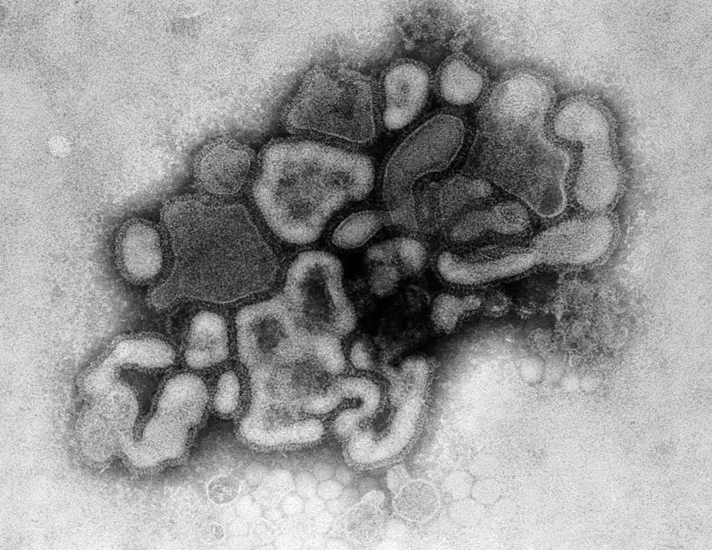
A sobering history lesson

The resurrection of an extinct but dangerous human-adapted H1N1 virus came about as the world was scrambling to prevent what was perceived to be the imminent emergence of a swine H1N1 influenza pandemic. People were so concerned about the possibility of a new pandemic that they inadvertently caused one. It was a self-fulfilling-prophecy pandemic.

I have no intent to lay blame here; indeed, my main point is that in the epidemiological fog of the moment in 1976, with anxiety mounting worldwide about a looming pandemic, a research unit in any country could have accidentally released the resurrected virus that came to be called the Russian flu. In the global rush to head off a possible new pandemic of H1N1 swine flu from Fort Dix through research and vaccination, accidents could have happened anywhere.

Advertisement

Of course, biocontainment facilities and policies have improved dramatically over the past half-century. But at the same time, there has been an equally dramatic proliferation of high-containment labs around the world.

woman fully contained in personal protective gear reaches across glass bottles

Across the globe, researchers work on dangerous pathogens in labs that are part of biocontainment facilities.

AP Photo/Michael Probst

Overreaction. Unintended consequences. Making matters worse. Self-fulfilling prophecy. There is a rich variety of terms to describe how the best intentions can go awry. Still reeling from , the world now faces new threats from cross-species jumps of avian flu viruses, mpox viruses and others. It’s critical that we be quick to respond to these emerging threats to prevent yet another global disease conflagration. Quick, but not too quick, history suggests.The Conversation

Donald S. Burke, Dean Emeritus and Distinguished University Professor Emeritus of Health Science and Policy, and of Epidemiology, at the School of Public Health, University of Pittsburgh

This article is republished from The Conversation under a Creative Commons license. Read the original article.

Advertisement

Read More

The post Preparing for a pandemic that never came ended up setting off another − how an accidental virus release triggered 1977’s ‘Russian flu’ appeared first on .com

The Conversation

Sunflowers make small moves to maximize their Sun exposure − physicists can model them to predict how they grow

Published

on

theconversation.com – Chantal Nguyen, Postdoctoral Associate at the BioFrontiers Institute, of Colorado Boulder – 2024-09-13 07:31:40

Sunflowers use tiny movements to follow the Sun’s path throughout the day.

AP Photo/Charlie Riedel

Chantal Nguyen, University of Colorado Boulder

Advertisement

Most of us aren’t spending our days watching our houseplants grow. We see their signs of only occasionally – a new leaf unfurled, a stem leaning toward the window.

But in the summer of 1863, Charles Darwin lay ill in bed, with nothing to do but watch his plants so closely that he could detect their small movements to and fro. The tendrils from his cucumber plants swept in circles until they encountered a stick, which they proceeded to twine around.

“I am getting very much amused by my tendrils,” he wrote.

This amusement blossomed into a decadeslong fascination with the little-noticed world of plant movements. He compiled his detailed observations and experiments in a 1880 book called “The Power of Movement in Plants.”

Advertisement

A zig-zagging line showing the movement of a leaf.

A diagram tracking the circumnutation of a leaf over three days.

Charles Darwin

In one study, he traced the motion of a carnation leaf every few hours over the course of three days, revealing an irregular looping, jagged path. The swoops of cucumber tendrils and the zags of carnation leaves are examples of inherent, ubiquitous plant movements called circumnutations – from the Latin circum, meaning circle, and nutare, meaning to nod.

Circumnutations vary in size, regularity and timescale across plant species. But their exact function remains unclear.

I’m a physicist interested in understanding collective behavior in living . Like Darwin, I’m captivated by circumnutations, since they may underlie more complex phenomena in groups of plants.

Advertisement

Sunflower patterns

A 2017 study revealed a fascinating observation that got my colleagues and me wondering about the role circumnutations could play in plant growth patterns. In this study, researchers found that sunflowers grown in a dense row naturally formed a near-perfect zigzag pattern, with each plant leaning away from the row in alternating directions.

This pattern the plants to avoid shade from their neighbors and maximize their exposure to sunlight. These sunflowers flourished.

Researchers then planted some plants at the same density but constrained them so that they could grow only upright without leaning. These constrained plants produced less oil than the plants that could lean and get the maximum amount of sun.

While farmers can’t grow their sunflowers quite this close together due to the potential for disease spread, in the future they may be able to use these patterns to up with new planting strategies.

Advertisement

Self-organization and randomness

This spontaneous pattern formation is a neat example of self-organization in nature. Self-organization refers to when initially disordered systems, such as a jungle of plants or a swarm of bees, achieve order without anything controlling them. Order emerges from the interactions between individual members of the system and their interactions with the .

Somewhat counterintuitively, noise – also called randomness – facilitates self-organization. Consider a colony of ants.

Ants secrete pheromones behind them as they crawl toward a food source. Other ants find this food source by the pheromone trails, and they further reinforce the trail they took by secreting their own pheromones in turn. Over time, the ants converge on the best path to the food, and a single trail prevails.

But if a shorter path were to become possible, the ants would not necessarily find this path just by following the existing trail.

Advertisement

If a few ants were to randomly deviate from the trail, though, they might stumble onto the shorter path and create a new trail. So this randomness injects a spontaneous change into the ants’ system that allows them to explore alternative scenarios.

Eventually, more ants would follow the new trail, and soon the shorter path would prevail. This randomness helps the ants adapt to changes in the environment, as a few ants spontaneously seek out more direct ways to their food source.

A group of honeybees spread out standing on honeycomb.

Beehives are an example of self-organization in nature.

Martin Ruegner/Stone via Getty Images

In biology, self-organized systems can be found at a range of scales, from the patterns of proteins inside cells to the socially complex colonies of honeybees that collectively build nests and forage for nectar.

Advertisement

Randomness in sunflower self-organization

So, could random, irregular circumnutations underpin the sunflowers’ self-organization?

My colleagues and I set out to explore this question by following the growth of young sunflowers we planted in the lab. Using cameras that imaged the plants every five minutes, we tracked the movement of the plants to see their circumnutatory paths.

We saw some loops and spirals, and lots of jagged movements. These ultimately appeared largely random, much like Darwin’s carnation. But when we placed the plants together in rows, they began to move away from one another, forming the same zigzag configurations that we’d seen in the previous study.

Five plants and a diagram showing loops and jagged lines that represent small movements made by the plants.

Tracking the circumnutations made by young sunflower plants.

Chantal Nguyen

Advertisement

We analyzed the plants’ circumnutations and found that at any given time, the direction of the plant’s motion appeared completely independent of how it was moving about half an hour earlier. If you measured a plant’s motion once every 30 minutes, it would appear to be moving in a completely random way.

We also measured how much the plant’s leaves grew over the course of two weeks. By putting all of these results together, we sketched a picture of how a plant moved and grew on its own. This information allowed us to computationally model a sunflower and simulate how it behaves over the course of its growth.

A sunflower model

We modeled each plant simply as a circular crown on a stem, with the crown expanding according to the growth rate we measured experimentally. The simulated plant moved in a completely random way, taking a “step” every half hour.

We created the model sunflowers with circumnutations of lower or higher intensity by tweaking the step sizes. At one end of the spectrum, sunflowers were much more likely to take tiny steps than big ones, leading to slow, minimal movement on average. At the other end were sunflowers that are equally as likely to take large steps as small steps, resulting in highly irregular movement. The real sunflowers we observed in our experiment were somewhere in the middle.

Advertisement

Plants require light to grow and have evolved the ability to detect shade and alter the direction of their growth in response.

We wanted our model sunflowers to do the same thing. So, we made it so that two plants that get too close to each other’s shade begin to lean away in opposite directions.

Finally, we wanted to see whether we could replicate the zigzag pattern we’d observed with the real sunflowers in our model.

First, we set the model sunflowers to make small circumnutations. Their shade avoidance responses pushed them away from each other, but that wasn’t enough to produce the zigzag – the model plants stayed stuck in a line. In physics, we would call this a “frustrated” system.

Advertisement

Then, we set the plants to make large circumnutations. The plants started moving in random patterns that often brought the plants closer together rather than farther apart. Again, no zigzag pattern like we’d seen in the field.

But when we set the model plants to make moderately large movements, similar to our experimental measurements, the plants could self-organize into a zigzag pattern that gave each sunflower optimal exposure to light.

So, we showed that these random, irregular movements helped the plants explore their surroundings to find desirable arrangements that benefited their growth.

Plants are much more dynamic than people give them credit for. By taking the time to follow them, scientists and farmers can unlock their secrets and use plants’ movement to their advantage.The Conversation

Chantal Nguyen, Postdoctoral Associate at the BioFrontiers Institute, University of Colorado Boulder

Advertisement

This article is republished from The Conversation under a Creative Commons license. Read the original article.

Read More

The post Sunflowers make small moves to maximize their Sun exposure − physicists can model them to predict how they grow appeared first on .com

Advertisement
Continue Reading

The Conversation

Endometriosis pain leads to missed school and work in two-thirds of women with the condition, new study finds

Published

on

theconversation.com – Rasha Al-Lami, Researcher in Women’s Health, Yale – 2024-09-13 07:30:43

Endometriosis affects about 10% of reproductive-age women worldwide.

Xavier Lorenzo/Moment via Getty Images

Rasha Al-Lami, Yale University

Advertisement

More than two-thirds of women with endometriosis missed school or work due to pain from the condition, in a study of more than 17,000 women between the ages of 15 and 44 in the U.S. That is a key finding of new research published in the Journal of Endometriosis and Uterine Disorders.

Our study also found that Black and Hispanic women were less likely to be diagnosed with endometriosis with white women. Interestingly, women who identified as part of the LGBTQ community had a higher likelihood of receiving an endometriosis diagnosis than heterosexual women.

We used data from the National Health and Nutrition Examination Survey, which is administered by the Centers for Disease Control and Prevention, for the period 2011 to 2019. The survey data use adjusted weights to account for the racial composition of U.S. society, meaning our sample of 17,619 women represents 51,981,323 women of the U.S. population.

We specifically examined factors related to quality of , such as poverty, education and functional impairment, as well as race and sexual orientation.

Advertisement

I am a physician-scientist and a researcher in women’s health, working together with specialists in OB-GYN from Yale and the University of .

Why it matters

Endometriosis is a chronic, often painful condition that affects approximately 10% of reproductive-age women worldwide. It occurs when tissues that would normally line the inner surface of the uterus instead occur outside the uterus, such as on the ovaries or even in distant organs such as the lungs or brain. These abnormally located lesions respond to hormonal changes during the menstrual cycle, causing pain when stimulated by the hormones that regulate the menstrual cycle.

Our study sheds light on how endometriosis, despite its prevalence, remains underdiagnosed and underresearched. We found that 6.4% of reproductive-age women in the U.S. had an endometriosis diagnosis. More than 67% reported missed work or school, or been unable to perform activities, due to pain associated with endometriosis.

Our study highlights disparities in the diagnosis and management of endometriosis among different racial groups. Black women had 63% lower odds of getting an endometriosis diagnosis, and Hispanic women had 55% lower odds compared with non-Hispanic white women. This disparity may reflect historical biases in , pointing to the need for more equitable practices.

Advertisement

In addition, our study underscores the importance of considering women’s health across diverse population subgroups, with particular attention to sexual orientation. We found that non-heterosexual lesbian, gay, bisexual, transgender and queer women had 54% higher odds of receiving an endometriosis diagnosis compared with straight women. Our study was the first to examine endometriosis likelihood among non-heterosexual women at the national level in the U.S.

We found no significant association between endometriosis and other quality-of-life indicators such as poverty, education or employment status, which suggests that the condition affects women across various socioeconomic backgrounds.

A common theory about the cause of endometriosis is that women have menstrual blood that seeds outside of the uterus, but recent research supports inflammatory causes.

What other research is being done

Our work adds to the growing body of evidence that Black women are less likely to be diagnosed with endometriosis and that their reported pain symptoms are often overlooked.

Explanations for this inequity include health care bias against minority women and limited access to medical care among Black women. Research also shows that many medical professionals as well as medical and residents believe that Black women have a lower pain threshold compared with the white population.

Advertisement

This is another possible reason that pain symptoms among Black women with endometriosis get neglected. Researchers from the U.K reported the same findings, attributing these disparities to systemic bias and inequitable medical care.

Another study estimates that the lifetime costs associated with having endometriosis are about US$27,855 per year per patient in the U.S., costing the country about $22 annually on health care expenditures.The Conversation

Rasha Al-Lami, Researcher in Women’s Health, Yale University

This article is republished from The Conversation under a Creative Commons license. Read the original article.

Read More

Advertisement

The post Endometriosis pain leads to missed school and work in two-thirds of women with the condition, new study finds appeared first on .com

Continue Reading

The Conversation

Biobots arise from the cells of dead organisms − pushing the boundaries of life, death and medicine

Published

on

theconversation.com – Peter A Noble, Affiliate Professor of Microbiology, University of Washington – 2024-09-12 07:31:37

Biobots could one day be engineered to deliver drugs and clear up arterial plaque.
Kriegman et al. 2020/PNAS, CC BY-SA

Peter A Noble, University of Washington and Alex Pozhitkov, Irell & Manella Graduate School of Biological Sciences at City of Hope

and death are traditionally viewed as opposites. But the emergence of new multicellular life-forms from the cells of a dead organism introduces a “third state” that lies beyond the traditional boundaries of life and death.

Usually, scientists consider death to be the irreversible halt of functioning of an organism as a whole. However, practices such as organ donation highlight how organs, tissues and cells can continue to function even after an organism’s demise. This resilience raises the question: What mechanisms allow certain cells to keep working after an organism has died?

Advertisement

We are researchers who investigate what happens within organisms after they die. In our recently published review, we describe how certain cells – when provided with nutrients, oxygen, bioelectricity or biochemical cues – have the capacity to transform into multicellular organisms with new functions after death.

Life, death and emergence of something new

The third challenges how scientists typically understand cell behavior. While caterpillars metamorphosing into butterflies, or tadpoles evolving into frogs, may be familiar developmental transformations, there are few instances where organisms change in ways that are not predetermined. Tumors, organoids and cell lines that can indefinitely divide in a petri dish, like HeLa cells, are not considered part of the third state because they do not develop new functions.

However, researchers found that skin cells extracted from deceased frog embryos were able to adapt to the new conditions of a petri dish in a lab, spontaneously reorganizing into multicellular organisms called xenobots. These organisms exhibited behaviors that extend far beyond their original biological roles. Specifically, these xenobots use their cilia – small, hair-like structures – to navigate and move through their surroundings, whereas in a living frog embryo, cilia are typically used to move mucus.

Xenobots can move, heal and interact with their environment on their own.

Xenobots are also able to perform kinematic self-replication, meaning they can physically replicate their structure and function without growing. This differs from more common replication processes that involve growth within or on the organism’s body.

Advertisement

Researchers have also found that solitary human lung cells can self-assemble into miniature multicellular organisms that can move around. These anthrobots behave and are structured in new ways. They are not only able to navigate their surroundings but also repair both themselves and injured neuron cells placed nearby.

Taken together, these findings demonstrate the inherent plasticity of cellular and challenge the idea that cells and organisms can evolve only in predetermined ways. The third state suggests that organismal death may play a significant role in how life transforms over time.

Microscopy images of a black blob fusing together two groundglass walls in three panels, and a green web plugging a gap in a web of pink
Diagram A shows an anthrobot building a bridge across a scratched neuron over the course of three days. Diagram B highlights the ‘stitch’ in green at the end of Day 3.
Gumuskaya et al. 2023/Advanced Science, CC BY-SA

Postmortem conditions

Several factors influence whether certain cells and tissues can survive and function after an organism dies. These include environmental conditions, metabolic activity and preservation techniques.

Different cell types have varying survival times. For example, in humans, white blood cells die between 60 and 86 hours after organismal death. In mice, skeletal muscle cells can be regrown after 14 days postmortem, while fibroblast cells from sheep and goats can be cultured up to a month or so postmortem.

Metabolic activity plays an important role in whether cells can continue to survive and function. Active cells that require a continuous and substantial supply of energy to maintain their function are more difficult to culture than cells with lower energy requirements. Preservation techniques such as cryopreservation can allow tissue samples such as bone marrow to function similarly to that of living donor sources.

Advertisement

Inherent survival mechanisms also play a key role in whether cells and tissues on. For example, researchers have observed a significant increase in the activity of stress-related genes and immune-related genes after organismal death, likely to compensate for the loss of homeostasis. Moreover, factors such as trauma, infection and the time elapsed since death significantly affect tissue and cell viability.

Microscopy image of developing white and red blood cells
Different cell types have different capacities for survival, white blood cells.
Ed Reschke/Stone via Getty Images

Factors such as age, , sex and type of species further shape the postmortem landscape. This is seen in the challenge of culturing and transplanting metabolically active islet cells, which produce insulin in the pancreas, from donors to recipients. Researchers believe that autoimmune processes, high energy costs and the degradation of protective mechanisms could be the reason behind many islet transplant failures.

How the interplay of these variables allows certain cells to continue functioning after an organism dies remains unclear. One hypothesis is that specialized channels and pumps embedded in the outer membranes of cells serve as intricate electrical circuits. These channels and pumps generate electrical that allow cells to communicate with each other and execute specific functions such as growth and movement, shaping the structure of the organism they form.

The extent to which different types of cells can undergo transformation after death is also uncertain. Previous research has found that specific genes involved in stress, immunity and epigenetic regulation are activated after death in mice, zebrafish and people, suggesting widespread potential for transformation among diverse cell types.

Implications for biology and medicine

The third state not only offers new insights into the adaptability of cells. It also offers prospects for new treatments.

Advertisement

For example, anthrobots could be sourced from an individual’s living tissue to deliver drugs without triggering an unwanted immune response. Engineered anthrobots injected into the body could potentially dissolve arterial plaque in atherosclerosis and remove excess mucus in cystic fibrosis patients.

Importantly, these multicellular organisms have a finite life span, naturally degrading after four to six weeks. This “kill switch” prevents the growth of potentially invasive cells.

A better understanding of how some cells continue to function and metamorphose into multicellular entities some time after an organism’s demise promise for advancing personalized and preventive medicine.The Conversation

Peter A Noble, Affiliate Professor of Microbiology, University of Washington and Alex Pozhitkov, Senior Technical of Bioinformatics, Irell & Manella Graduate School of Biological Sciences at City of Hope

This article is republished from The Conversation under a Creative Commons license. Read the original article.

Advertisement

Read More

The post Biobots arise from the cells of dead organisms − pushing the boundaries of life, death and medicine appeared first on theconversation.com

Continue Reading

Trending