Connect with us

The Conversation

Fluid keeps your brain from crushing itself and shields your spine from shock – a neurologist explains what happens when it stops working

Published

on

theconversation.com – Danielle Wilhour, Assistant Professor of Neurology, University of Colorado Anschutz Medical Campus – 2024-08-14 07:30:04

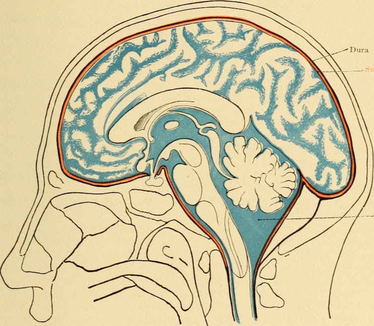
Your brain produces around a pint of cerebrospinal fluid each day.
Archibald Church, Diseases of the nervous system (1908)/Internet Archive via Flickr

Danielle Wilhour, University of Colorado Anschutz Medical Campus

Cerebrospinal fluid, or CSF, is a clear, colorless liquid that plays a crucial role in maintaining the health and function of your central nervous system. It cushions the brain and spinal cord, provides nutrients and removes waste products.

Despite its importance, problems related to CSF often go unnoticed until something goes wrong.

I am a neurologist and headache specialist. In my work treating patients with CSF pressure disorders, I have seen these conditions present in many different ways. Here’s what happens when your cerebrospinal fluid stops working:

What is cerebrospinal fluid?

CBF is made of water, proteins, sugar, ions and neurotransmitters. It is primarily produced by a network of cells called the choroid plexus, which is located in the brain’s ventricles, or cavities.

The choroid plexus produces approximately 500 milliliters of CSF daily, but only about 150 milliliters are present within the central nervous system at any given time due to constant absorption and replenishment in the brain. This fluid circulates through the ventricles of the brain, the central canal of the spinal cord and the subarachnoid space surrounding the brain and spinal cord.
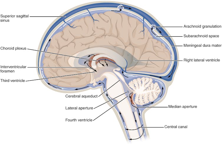
Diagram of cross-section of brain depicting cerebrospinal fluid circulation
Cerebrospinal fluid circulates throughout the brain and spinal cord.
OpenStax, CC BY-SA

CSF has several critical functions. It protects the brain and spinal cord from injury by absorbing shocks. Suspending the brain in this fluid reduces its effective weight and prevents it from being crushed under its own mass. Additionally, CSF helps maintain a stable chemical environment in the central nervous system, facilitating the removal of metabolic waste and the distribution of nutrients and hormones.

When the production, circulation or absorption of cerebrospinal fluid is disrupted, this can lead to significant health issues. Two notable conditions are CSF leaks and idiopathic intracranial hypertension.

CSF leak

A CSF leak occurs when the fluid escapes through a tear or hole in the dura mater – the tough, outermost layer of the meninges that surrounds the brain and spinal cord.

The dura can be damaged from head injuries or punctured during surgical procedures involving the sinuses, brain or spine, such as lumbar puncture, epidurals, spinal anesthesia or myelogram. Spontaneous CSF leaks can also occur without any identifiable cause.

CSF leaks were originally thought to be relatively rare, with an estimated annual incidence of 5 per 100,000 people. However, with increased awareness and advances in imaging, health care providers are discovering more and more leaks. They tend to occur more frequently in middle-aged adults and are more common in women than men.

Risk factors for the condition include connective tissue disorders such as Ehlers-Danlos syndrome as well as postural orthostatic tachycardia syndrome.

An upright headache could be a sign of a CSF leak.

Unfortunately, it’s common for health care providers to misdiagnose a CSF leak as another condition, like migraine, sinus infections or allergies. What can make diagnosing a CSF leak challenging is its broad symptoms. Most people with CSF leak have a positional headache that improves when lying down and worsens when standing. Pain is usually felt in the back of the head and may involve the neck and between the shoulder blades. In addition to headaches, patients may experience ringing in the ears, vision disturbances, memory problems, brain fog, dizziness and nausea.

Imaging may help guide diagnosis, including an MRI of your brain or entire spine, or a myelogram of the space surrounding your spinal cord. Features of a CSF leak that are visible in a scan include your brain sagging down in the base of your skull as well as a fluid collection outside of your dura. However, an estimated 19% of people with a CSF leak can have normal scans, so not seeing signs of a leak on imaging does not entirely rule it out.

Conservative treatment for a CSF leak involves rest, lying flat and increasing your fluid intake to give your spine time to heal the puncture. Increasing your caffeine consumption to an equivalent of three to four cups of coffee per day can also help by increasing CSF production through stimulating the choroid plexus. Caffeine also relieves pain by interacting with adenosine receptors, which are key players in the body’s pain perception mechanisms.

If a conservative approach is not successful, an epidural blood patch may be necessary. In this procedure, blood is drawn from your arm and injected into your spine. The injected blood can help form a covering over the hole and promote the healing process. Headache improvement can be fast, but if the patch does not work or the results are short-lived, additional testing may be needed to better locate the site of the leak. In rare cases, surgery may be recommended. Most patients with CSF leak respond to some form of these treatments.

Idiopathic intracranial hypertension

Idiopathic intracranial hypertension is a disorder involving an excess of CSF that elevates pressure inside the skull and compresses the brain. The term “idiopathic” indicates that the cause of the raised pressure is unknown.

Most patients with idiopathic intracranial hypertension have a history of obesity or recent weight gain. Other risk factors include taking certain medications such as tetracycline, excessive vitamin A, tretinoin, steroids and growth hormone. Middle-aged obese women are 20 times more likely to be diagnosed with idiopathic intracranial hypertension than other patient groups. As obesity becomes more prevalent, so too does the incidence of this condition.

Idiopathic intracranial hypertension results from increased intracranial pressure.

Patients with idiopathic intracranial hypertension typically experience headaches and vision changes, tinnitus or eye pain. Papilledema, or swelling of the optic disc, is the hallmark finding on a fundoscopic examination of the back of the eye. Clinicians may also observe paralysis of the patient’s eye muscles.

Brain imaging of patients suspected of having idiopathic intracranial hypertension is crucial to excluding other causes of elevated CSF pressure, such as brain tumors or blood clots in the brain. A lumbar puncture or spinal tap to measure the pressure and composition of CSF is also central to diagnosis.

Since high intracranial pressure can damage the optic nerve and lead to permanent vision loss, the primary goal of treatment is to decrease pressure and preserve the optic nerve. Treatment options include weight loss, dietary changes and medications to reduce CSF production. Surgical procedures can also reduce intracranial pressure.

Future directions and unknowns

Cerebrospinal fluid is indispensable for brain health. Despite advancements in understanding diseases related to CSF, several aspects remain unclear.

The exact mechanisms that lead to conditions like CSF leaks and idiopathic intracranial hypertension are not fully understood, though there are many theories. Further research is vital to enhance diagnostic accuracy and effective treatments for CSF disorders.The Conversation

Danielle Wilhour, Assistant Professor of Neurology, University of Colorado Anschutz Medical Campus

This article is republished from The Conversation under a Creative Commons license. Read the original article.

Read More

The post Fluid keeps your brain from crushing itself and shields your spine from shock – a neurologist explains what happens when it stops working appeared first on theconversation.com

The Conversation

As the Taurid meteor shower passes by Earth, pseudoscience rains down – and obscures a potential real threat from space

Published

on

theconversation.com – Mark Boslough, Research Associate Professor of Earth and Planetary Sciences, University of New Mexico – 2024-11-15 07:31:00

This image of a Taurid fireball was taken in 2014 by NASA’s All Sky Fireball Network in Tullahoma, Tenn.

NASA

Mark Boslough, University of New Mexico

With the Taurid meteor shower now hitting the night skies worldwide, look for what could be a celestial treat – you might see shooting stars, and maybe even fireballs, the biggest and brightest meteors.

As the full moon begins to wane after Nov. 15, the sky will be darker, due to diminishing moonlight, so finding the meteors will get easier. That said, the best visibility for the meteors through the rest of the month will come just before moonrise each night.

Beyond the light show, there is something else that scientists as well as onlookers have long wondered about: the possibility that bigger chunks are in the Taurid meteor streams, chunks the size of boulders, buildings or even mountains.

And if that’s true, could one of those monster-sized Taurid objects collide with Earth? Could they wipe out a city, or worse? Is it possible that’s already happened, sometime in our planet’s past?

This animation simulates the motion of the hypothetical Taurid meteor swarm through space.

As a physicist who researches the risk that comets and asteroids pose to the Earth, I’m aware that this is a subject where pseudoscience often competes with actual science. So let’s try to find the line between fact and fiction.

Pig Pen, glowing tails and shooting stars

Comet Encke is the so-called parent comet of the Taurid meteors. It’s relatively small, just over 3 miles (almost 5 kilometers) in diameter, and crosses inside Earth’s orbit and back out every 3.3 years.

As Encke moves, it sheds dust wherever it goes, like the Peanuts character Pig Pen. A meteor shower occurs when that dust and debris light up while entering Earth’s atmosphere at high speeds. Ultimately, they vanish into an incandescent puff of vapor with a glowing tail, creating the illusion of a “shooting star.”

But dust isn’t all that breaks off the comet. So do bigger chunks, the size of pebbles and stones. When they collide with the air, they create the much brighter fireballs, which sometimes explode.

Against a black and white starscape, a bright spot appears in the center of the photo.

An image of comet Encke, taken by NASA’s MESSENGER spacecraft in November 2013.

NASA/Johns Hopkins University Applied Physics Laboratory/Carnegie Institution of Washington/Southwest Research Institute

Doomsday showers

The “coherent catastrophism” hypothesis suggests that comet Encke was created when an even larger comet broke up into pieces; Encke survived as the largest piece. The hypothesis also suggests that other mountain-sized chunks broke off and coalesced into a large swarm of fragments too. If such a swarm exists, there is a possibility that those large chunks could one day hit Earth as it passes through the swarm.

But just because something might be physically possible doesn’t mean that it exists. Mainstream astronomers have rejected this theory’s most catastrophic predictions. Among other reasons, scientists have never observed high concentrations of these mountain-sized objects.

Despite the lack of evidence, researchers on the fringes of science have embraced the idea. They claim the Earth experienced a global catastrophic swarm 12,900 years ago; the impact, they say, caused continent-wide firestorms, floods and abrupt climate change that led to the mass extinction of large mammals, such as woolly mammoths, and the disappearance of early Americans known as the Clovis people.

The evidence for a catastrophic cause of these events, most of which did not happen, is lacking. Nevertheless, the idea has gained a large following and formed the basis for British author Graham Hancock’s popular TV series, “Ancient Apocalypse.”

A black and white photo of a forest of flattened trees.

This photo shows the flattened trees resulting from the Tunguska event.

Universal History Archives/Universal Images Group via Getty Images

The Tunguska event

But even outlandish ideas can have elements of truth, and there are hints that some objects – more than just dust and debris, but less than doomsday size – indeed exist in the Taurid meteor stream, and that the Earth has already encountered them.

One clue comes from an event on June 30, 1908, when an enormous explosion in the sky blew down millions of trees in Siberia. This was the Tunguska event – an airburst from an object that may have been up to 160 feet (about 50 meters) in diameter.

The collision unleashed several megatons of energy, which is roughly the equivalent of a large thermonuclear bomb. What happens is this: The incoming object penetrates deep into Earth’s atmosphere, and the dense air slows it down and heats it up until it vaporizes and explodes.

Could this object have been a Taurid? After all, the Taurids cross Earth’s orbit twice a year – not just in autumn, but also in June.

A fireball appears in the night sky.

In a 2015 photo, a glowing Taurid fireball descends over Lake Simcoe in Ontario, Canada.

Orchidpost/iStock via Getty Images Plus

Here’s the evidence: First, the descriptions of the trajectory of the Tunguska airburst, as reported by eyewitness observers, is consistent with that of an object coming from the Taurid stream.

What’s more, the pattern of blast damage on the ground beneath an airburst depends on the trajectory of the exploding object. Supercomputer simulations show that the shape of the surface blast that would be caused by an exploding Taurid object matches the pattern of fallen trees at Tunguska.

Finally, during the Taurid meteor shower in 1975, people observed large fireballs – and seismometers, previously placed on the Moon by Apollo astronauts, detected seismic events on the lunar surface. Scientists interpreted those events as impacts, presumably made by the Taurid meteors.

In 2032 and 2036, the Taurid swarm – assuming it exists – is predicted to be closer to the Earth than any time since 1975. That might mean the Moon, and perhaps the Earth, could be pelted again in those years.

There is time to figure this out. Scientists can expand their astronomical surveys to look for Tunguska-sized objects at the locations where they are predicted to be the next time they are in our vicinity.

Most scientists remain skeptical that such a swarm exists, but it’s the job of planetary defenders to investigate possible threats, even if the risk is small. After all, a Tunguska-sized object could conceivably demolish a major city and kill millions; an accurate count of objects on a potential collision course is essential.

Put doomsday scenarios and ancient apocalypses aside. The real question, and still an open one, is whether a Taurid swarm could deliver more Tunguska-sized objects than would otherwise be expected. This would mean we have underestimated the risk from future airbursts.The Conversation

Mark Boslough, Research Associate Professor of Earth and Planetary Sciences, University of New Mexico

This article is republished from The Conversation under a Creative Commons license. Read the original article.

Read More

The post As the Taurid meteor shower passes by Earth, pseudoscience rains down – and obscures a potential real threat from space appeared first on theconversation.com

Continue Reading

The Conversation

Knee problems tend to flare up as you age – an orthopedic specialist explains available treatment options

Published

on

theconversation.com – Angie Brown, Clinical Associate Professor of Physical Therapy, Quinnipiac University – 2024-11-15 07:32:00

Knee problems can hinder mobility and erode your quality of life.

Witthaya Prasongsin/Moment via Getty Images

Angie Brown, Quinnipiac University

Knee injuries are common in athletes, accounting for 41% of all athletic injuries. But knee injuries aren’t limited to competitive athletes. In our everyday lives, an accident or a quick movement in the wrong direction can injure the knee and require medical treatment. A quarter of the adult population worldwide experiences knee pain each year

As a physical therapist and board-certified orthopedic specialist, I help patients of all ages with knee injuries and degenerative conditions.

Your knees have a huge impact on your mobility and overall quality of life, so it’s important to prevent knee problems whenever possible and address pain in these joints with appropriate treatments.

Healthy knees

The knee joint bones consist of the femur, tibia and patella. As in all healthy joints, smooth cartilage covers the surfaces of the bones, forming the joints and allowing for controlled movement.

diagram of a healthy knee

A healthy knee.

Inna Kharlamova/iStock/Getty Images Plus via Getty Images

Muscles, ligaments and tendons further support the knee joint. The anterior cruciate ligament, commonly known as the ACL, and posterior cruciate ligament, or PCL, provide internal stability to the knee. In addition, two tough pieces of fibrocartilage, called menisci, lie inside the joint, providing further stability and shock absorption.

All these structures work together to enable the knee to move smoothly and painlessly throughout everyday movement, whether bending to pick up the family cat or going for a run.

Causes of knee pain

Two major causes of knee pain are acute injury and osteoarthritis.

Ligaments such as the ACL and PCL can be stressed and torn when a shear force occurs between the femur and tibia. ACL injuries often occur when athletes land awkwardly on the knee or quickly pivot on a planted foot. Depending on the severity of the injury, these patients may undergo physical therapy, or they may require surgery for repair or replacement.

PCL injuries are less common. They occur when the tibia experiences a posterior or backward force. This type of injury is common in car accidents when the knee hits the dashboard, or when patients fall forward when walking up stairs.

The menisci can also experience degeneration and tearing from shear and rotary forces, especially during weight-bearing activities. These types of injuries often require rehabilitation through physical therapy or surgery.

Knee pain can also result from injury or overuse of the muscles and tendons surrounding the knee, including the quadriceps, hamstrings and patella tendon.

Both injuries to and overuse of the knee can lead to degenerative changes in the joint surfaces, known as osteoarthritis. Osteoarthritis is a progressive disease that can lead to pain, swelling and stiffness. This disease affects the knees of over 300 million people worldwide, most often those 50 years of age and up. American adults have a 40% chance of developing osteoarthritis that affects their daily lives, with the knee being the most commonly affected joint.

Age is also a factor in knee pain. The structure and function of your joints change as you age. Cartilage starts to break down, your body produces less synovial fluid to lubricate your joints, and muscle strength and flexibility decrease. This can lead to painful, restricted movement in the joint.

Risk factors

There are some risk factors for knee osteoarthritis that you cannot control, such as genetics, age, sex and your history of prior injuries.

Fortunately, there are several risk factors you can control that can predispose you to knee pain and osteoarthritis specifically. The first is excessive weight. Based on studies between 2017 and 2020, nearly 42% of all adult Americans are obese. This obesity is a significant risk factor for diabetes and osteoarthritis and can also play a role in other knee injuries.

A lack of physical activity is another risk, with 1 in 5 U.S. adults reporting that they’re inactive outside of work duties. This can result in less muscular support for the knee and more pressure on the joint itself.

An inflammatory diet also adds to the risk of knee pain from osteoarthritis. Research shows that the average American diet, often high in sugar and fat and low in fiber, can lead to changes to the gut microbiome that contribute to osteoarthritis pain and inflammation.

Preventing knee pain

Increasing physical activity is one of the key elements to preventing knee pain. Often physical therapy intervention for patients with knee osteoarthritis focuses on strengthening the knee to decrease pain and support the joint during movement.

The U.S. Department of Health and Human Services recommends that adults spend at least 150 to 300 minutes per week on moderate-intensity, or 75 to 150 minutes per week on vigorous-intensity aerobic physical activity. These guidelines do not change for adults who already have osteoarthritis, although their exercise may require less weight-bearing activities, such as swimming, biking or walking.

The agency also recommends that all adults do some form of resistance training at least two or more days a week. Adults with knee osteoarthritis particularly benefit from quadriceps-strengthening exercises, such as straight leg raises.

Treatments for knee pain

Conservative treatment of knee pain includes anti-inflammatory and pain medications and physical therapy.

Medical treatment for knee osteoarthritis may include cortisone injections to decrease inflammation or hyaluronic acid injections, which help lubricate the joint. The relief from these interventions is often temporary, as they do not stop the progression of the disease. But they can delay the need for surgery by one to three years on average, depending on the number of injections.

Physical therapy is generally a longer-lasting treatment option for knee pain. Physical therapy treatment leads to more sustained pain reduction and functional improvements when compared with cortisone injections treatment and some meniscal repairs.

Patients with osteoarthritis often benefit from total knee replacement, a surgery with a high success rate and lasting results.

Surgical interventions for knee pain include the repair, replacement or removal of the ACL, PCL, menisci or cartilage. When more conservative approaches fail, patients with osteoarthritis may benefit from a partial or total knee replacement to allow more pain-free movement. In these procedures, one or both sides of the knee joint are replaced by either plastic or metal components. Afterward, patients attend physical therapy to aid in the return of range of motion.

Although there are risks with any surgery, most patients who undergo knee replacement benefit from decreased pain and increased function, with 90% of all replacements lasting more than 15 years. But not all patients are candidates for such surgeries, as a successful outcome depends on the patient’s overall health and well-being.

New treatments on the horizon

New developments for knee osteoarthritis are focused on less invasive therapies. Recently, the U.S. Food and Drug Administration approved a new implant that acts as a shock absorber. This requires a much simpler procedure than a total knee replacement.

Other promising interventions include knee embolization, a procedure in which tiny particles are injected into the arteries near the knee to decrease blood flow to the area and reduce inflammation near the joint. Researchers are also looking into injectable solutions derived from human bodies, such as plasma-rich protein and fat cells, to decrease inflammation and pain from osteoarthritis. Human stem cells and their growth factors also show potential in treating knee osteoarthritis by potentially improving muscle atrophy and repairing cartilage.

Further research is needed on these novel interventions. However, any intervention that holds promise to stop or delay osteoarthritis is certainly encouraging for the millions of people afflicted with this disease.The Conversation

Angie Brown, Clinical Associate Professor of Physical Therapy, Quinnipiac University

This article is republished from The Conversation under a Creative Commons license. Read the original article.

Read More

The post Knee problems tend to flare up as you age – an orthopedic specialist explains available treatment options appeared first on theconversation.com

Continue Reading

The Conversation

Get chronic UTIs? Future treatments may add more bacteria to your bladder to beat back harmful microbes

Published

on

theconversation.com – Sarguru Subash, Assistant Professor of Veterinary Pathobiology, Texas A&M University – 2024-11-15 07:31:00

Certain strains of E. coli can outcompete disease-causing microbes for resources.

NIAID/Flickr, CC BY-SA

Sarguru Subash, Texas A&M University

Millions of people in the U.S. and around the world suffer from urinary tract infections every year. Some groups are especially prone to chronic UTIs, including women, older adults and some veterans.

These infections are typically treated with antibiotics, but overusing these drugs can make the microbes they target become resistant and reduce the medicines’ effectiveness.

To solve this problem of chronic UTIs and antibiotic resistance, we combined our expertise in microbiology and engineering to create a living material that houses a specific strain of beneficial E. coli. Our research shows that the “good” bacteria released from this biomaterial can compete with “bad” bacteria for nutrients and win, dramatically reducing the number of disease-causing microbes.

With further development, we believe this technique could help manage recurring UTIs that do not respond to antibiotics.

Bringing bacteria to the bladder

For the microbes living in people, nutrients are limited their presence varies between different parts of the body. Bacteria have to compete with other microbes and the host to acquire essential nutrients. By taking up available nutrients, beneficial bacteria can stop or slow the growth of harmful bacteria. When harmful bacteria are starved of important nutrients, they aren’t able to reach high enough numbers to cause disease.

Delivering beneficial bacteria to the bladder to prevent UTIs in challenging, though. For one, these helpful bacteria can naturally colonize only in people who are unable to fully empty their bladder, a condition called urinary retention. Even among these patients, how long these bacteria can colonize their bladders varies widely.

Current methods to deliver bacteria to the bladder are invasive and require repeated catheter insertion. Even when bacteria are successfully released into the bladder, urine will flush out these microbes because they cannot stick to the bladder wall.

Micrograph of clusters of pink rods scattered across a blue textured wall, while yellow blobs extend thin tendrils across the clumps

This microscopy image shows the bladder of a mouse (blue) covered with E. coli (pink) and the white blood cells (yellow) attacking them.

Valerie O’Brien, Matthew Joens, Scott J. Hultgren, James A.J. Fitzpatrick, Washington University, St. Louis/NIH via Flickr, CC BY-NC

Biomaterials to treat UTIs

Since beneficial bacteria cannot attach to and survive in the bladder for long, we developed a biomaterial that could slowly release bacteria in the bladder over time.

Our biomaterial is composed of living E. coli embedded in a matrix structure made of gel. It resembles a piece of jelly about 500 times smaller than a drop of water and can release bacteria for up to two weeks in the bladder. By delivering the bacteria via biomaterial, we overcome the need for the bacteria to attach to the bladder to persist in the organ.

We tested our biomaterial by placing it in human urine in petri dishes and exposing it to bacterial pathogens that cause UTIs. Our results showed that when mixed in a 50:50 ratio, the E. coli outcompeted the UTI-causing bacteria by increasing to around 85% of the total population. When we added more E. coli than UTI-causing bacteria, which is what we envision for future development and testing, the proportion of E. coli increased to over 99% of the population, essentially wiping out the UTI-causing bacteria. Moreoever, the biomaterial continued releasing E. coli for up to two weeks in human urine.

Our findings suggest that E.coli could stick around and survive in the bladder for extended periods of time and successfully decrease the growth of many types of bacteria that cause UTIs.

Person pressing hands against stomach

UTIs can be painful.

Images we create and what actually happens are always beautiful when we have imagination/iStock via Getty Images Plus

Improving biomaterials

Our findings show that E. coli can not only control harmful bacteria it’s closely related to but also a broad range of disease-causing bacteria in humans and animals. This means scientists might not need to identify different types of beneficial bacteria to control each pathogen – and there are many – that can cause a UTI.

Our team is currently evaluating how effectively our biomaterial can cure UTIs in mice. We are also working to identify the specific nutrients that beneficial and harmful bacteria compete over and what factors may help beneficial bacteria win. We could add these nutrients to our biomaterial to be released or withheld.

This research is still at an early stage, and clinical uses are not in development yet, so if it does reach patients it will be well in the future. We hope that our technology could be refined and applied to control other bacterial infections and some cancers caused by bacteria.The Conversation

Sarguru Subash, Assistant Professor of Veterinary Pathobiology, Texas A&M University

This article is republished from The Conversation under a Creative Commons license. Read the original article.

Read More

The post Get chronic UTIs? Future treatments may add more bacteria to your bladder to beat back harmful microbes appeared first on theconversation.com

Continue Reading

Trending